- Home
- About ANT
-
Products
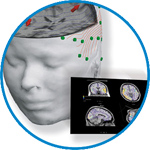
asa
asa is a highly flexible EEG/ERP and MEG analysis package with a variety of source reconstruction, signal analysis and MRI processing features.
.jpg)
eego mylab
The new frontier in multimodal brain research. With up to 16 kHz sampling rate, 256 EEG channels and unique software features, eego mylab gives you an unprecedented in-depth understanding of the human brain.
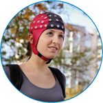
eego sports
eego sports offers complete freedom to collect high-density EEG data, bipolar EMG signals, and a variety of physiological sensor data, wherever and whenever required, with publish quality data in less than 15 minutes!
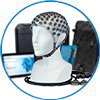
waveguard net
The waveguard net sets a new standard for research applications requiring high-density EEG data acquisition with quick preparation time, high flexibility, and subject comfort.
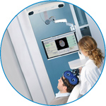
visor2
Our new and upgraded visor2 solutions integrate all the latest technologies for navigated rTMS, dual-coil navigation support, EEG-TMS recordings and pre-surgical evaluation for the highest quality in research and clinical procedures.
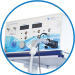
powerMAG ANT
The PowerMAG ANT 100 rTMS stimulator is designed for the specific needs of high-end TMS applications. Powerful high-frequency TMS as well as high precise single pulse and repetitive pulse protocols are combined in one single device.
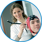
xensor
xensor offers the solution for digitization of 3D electrode positions. xensor takes care of the whole procedure; it records, visualizes and stores positions acquired with a dedicated digitizer.
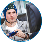
waveguard original
waveguard original is the cap solution for EEG measurements compatible with fMRI, MEG and TMS system. Use of active shielding guarantees performance in even the most demanding environments.
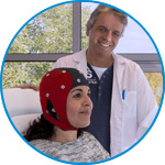
waveguard connect
waveguard connect EEG caps are a perfect match for hospitals and institutes aiming at reliable EEG, maximum uptime and great patient comfort! For optimal signal quality, the electrodes are made of pure, solid tin.
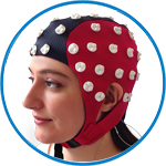
waveguard touch
waveguard touch is a dry electrode EEG cap. The unique Ag/AgCl coated soft polymer electrodes provide stable, research-grade EEG signals while maintaining subject comfort. The combination of these innovative dry electrodes and the industry-leading waveguard cap makes waveguard touch the best solution for dry EEG.
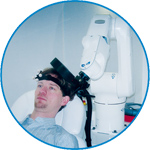
smartmove
smartmove allows planning of a complete TMS session ahead by defining stimulation sites based on anatomical MRI information and functional information like fMRI, PET or EEG/MEG.
Stay - References
- Support
- Events
- News
- Contact Us
You are here
Timing of V1/V2 and V5+ activations during coherent motion of dots: An MEG study
Timing of V1/V2 and V5+ activations during coherent motion of dots: An MEG study
In order to study the temporal activation course of visual areas V1 and V5 in response to a motion stimulus, a random dots kinematogram paradigm was applied to eight subjects while magnetic fields were recorded using magnetoencephalography (MEG). Sources generating the registered magnetic fields were localized with Magnetic Field Tomography (MFT). Anatomical identification of cytoarchitectonically defined areas V1/V2 and V5 was achieved by means of probabilistic cytoarchitectonic maps. We found that the areas V1/V2 and V5+ (V5 and other adjacent motion sensitive areas) exhibited two main activations peaks at 100–130 ms and at 140–200 ms after motion onset. The first peak found for V1/V2, which corresponds to the visual evoked field (VEF) M1, always preceded the peak found in V5+. Additionally, the V5+ peak was correlated significantly and positively with the second V1/V2 peak. This result supports the idea that the M1 component is generated not only by the visual area V1/V2 (as it is usually proposed), but also by V5+. It reflects a forward connection between both structures, and a feedback projection to V1/V2, which provokes a second activation in V1/V2 around 200 ms. This second V1/V2activation (corresponding to motion VEF M2) appeared earlier than the second V5+ activation but both peaked simultaneously. This result supports the hypothesis that both areas also generate the M2 component, which reflects a feedback input from V5+ to V1/V2 and a crosstalk between both structures. Our study indicates that during visual motion analysis, V1/V2 and V5+ are activated repeatedly through forward and feedback connections and both contribute to m-VEFs M1 and M2.

 Read more
.jpg)


