- Home
- About ANT
-
Products
asa
asa is a highly flexible EEG/ERP and MEG analysis package with a variety of source reconstruction, signal analysis and MRI processing features.
.jpg)
eego mylab
The new frontier in multimodal brain research. With up to 16 kHz sampling rate, 256 EEG channels and unique software features, eego mylab gives you an unprecedented in-depth understanding of the human brain.
eego sports
eego sports offers complete freedom to collect high-density EEG data, bipolar EMG signals, and a variety of physiological sensor data, wherever and whenever required, with publish quality data in less than 15 minutes!
waveguard net
The waveguard net sets a new standard for research applications requiring high-density EEG data acquisition with quick preparation time, high flexibility, and subject comfort.
visor2
Our new and upgraded visor2 solutions integrate all the latest technologies for navigated rTMS, dual-coil navigation support, EEG-TMS recordings and pre-surgical evaluation for the highest quality in research and clinical procedures.
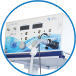
powerMAG ANT
The PowerMAG ANT 100 rTMS stimulator is designed for the specific needs of high-end TMS applications. Powerful high-frequency TMS as well as high precise single pulse and repetitive pulse protocols are combined in one single device.
xensor
xensor offers the solution for digitization of 3D electrode positions. xensor takes care of the whole procedure; it records, visualizes and stores positions acquired with a dedicated digitizer.
waveguard original
waveguard original is the cap solution for EEG measurements compatible with fMRI, MEG and TMS system. Use of active shielding guarantees performance in even the most demanding environments.
waveguard connect
waveguard connect EEG caps are a perfect match for hospitals and institutes aiming at reliable EEG, maximum uptime and great patient comfort! For optimal signal quality, the electrodes are made of pure, solid tin.
waveguard touch
waveguard touch is a dry electrode EEG cap. The unique Ag/AgCl coated soft polymer electrodes provide stable, research-grade EEG signals while maintaining subject comfort. The combination of these innovative dry electrodes and the industry-leading waveguard cap makes waveguard touch the best solution for dry EEG.
smartmove
smartmove allows planning of a complete TMS session ahead by defining stimulation sites based on anatomical MRI information and functional information like fMRI, PET or EEG/MEG.
- References
- Support
- Events
- News
- Contact Us
 Read more
.jpg)
You are here
Comparison of waveguard touch dry electrodes to conventional gelled electrodes on physiological paradigms
Comparison of waveguard touch dry electrodes to conventional gelled electrodes on physiological paradigms

|

|
Introduction
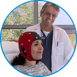
Professor David Liley and Dr. Levin Kuhlmann from the Centre for Human Psychopharmacology at Swinburne University of Technology have been working on combining MEG and EEG recordings to optimize treatment for patients suffering from epilepsy. They are partners in a collaborative study with one of the largest adult surgical epilepsy programs in Australia (SVHM) and aim for faster patient preparation, cap application, and electrode digitization, which are much needed for high-density intraoperative EEG recording in epileptic subjects.
A multicentric program has been conducted to evaluate and compare the dry electrode signal quality offered by the waveguard touch to the conventional gelled Ag/AgCl sensors.
Relevant physiological paradigms such as eye blinks, checkerboard pattern reversal visual evoked potential (VEP) occurrence, morphology and topography as well as resting state EEG spectra were extracted and examined.
Method
In this study, high density EEGs of 20 healthy adults (10/10 male/female) were recorded. The population group ranged from 23 to 60 yrs. old and were free from psychiatric/neurological issues or skin diseases. None of the participants were wearing make-up and all subjects had a head circumference measuring between 53.5 and 59.7 cm. All participants washed and dried their hair prior to the first electrode recording. EEGs were sequentially recorded with:
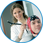
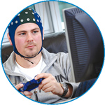
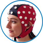
- A 64-channel waveguard touch dry electrode cap
- A 64-channel waveguard original conventional cap
- A 64-channel eego amplifier
For each participant, dry and gelled recordings were acquired sequentially, starting with 5 min resting EEG (eyes closed and eyes open), 20 eye blinks for 40 seconds, and pattern reversal VEP according to ISCEV 2010 standard.
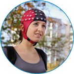
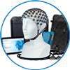
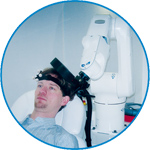
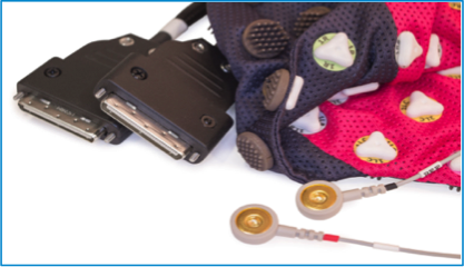
Figure 1 and Figure 2 show the setup of the experiment and the waveguard touch that was used in this experiment.
|
|
EEG Data
Eye blinks and resting EEG
Figure 3 displays the eye blinks recorded in two typical patients using dry and conventional gelled electrodes. Blue indicates the conventional cap, whilst red illustrates the dry electrode cap.
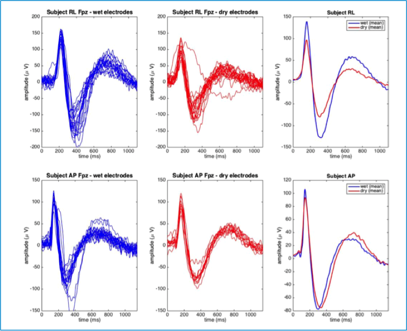
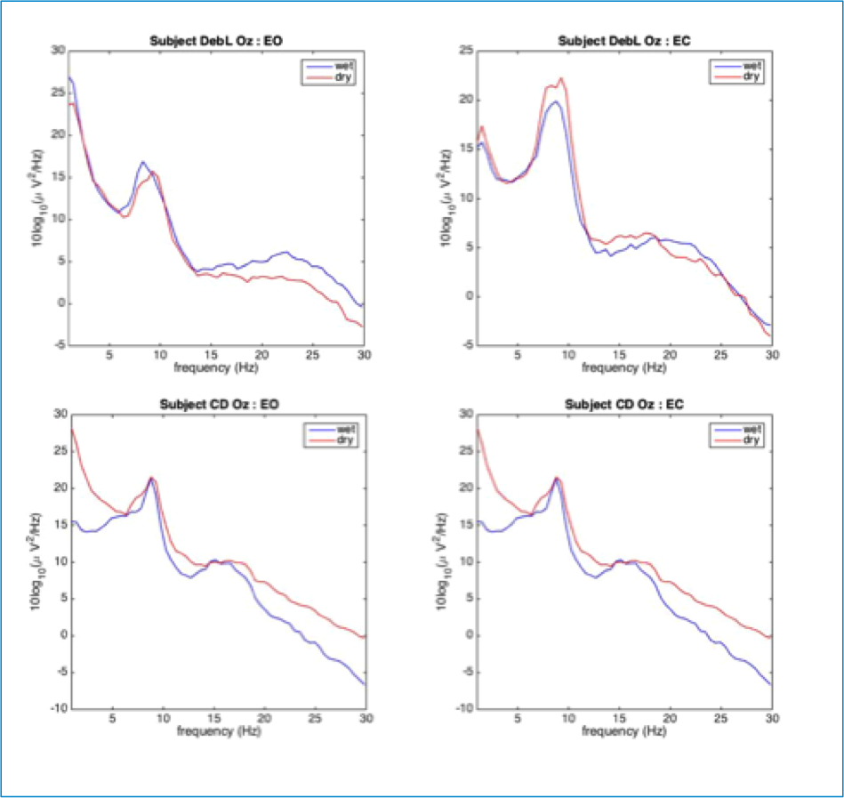
Figure 4 shows the resting state EEGs of two typical subjects in the frequency domain. The increase of alpha band during eyes closed is prominent in the first subject. The overlay of gelled and dry recordings shows the same physiologies for each subject during both the eyes open and eyes closed sessions.
VEP morphology and VEP topography
The dry and gelled recordings were also compared on their performance regarding VEP morphology and topography. As shown in the overlay plot in Figure 5 and in Figure 6, the waveforms for dry and gelled electrodes were qualitatively similar.
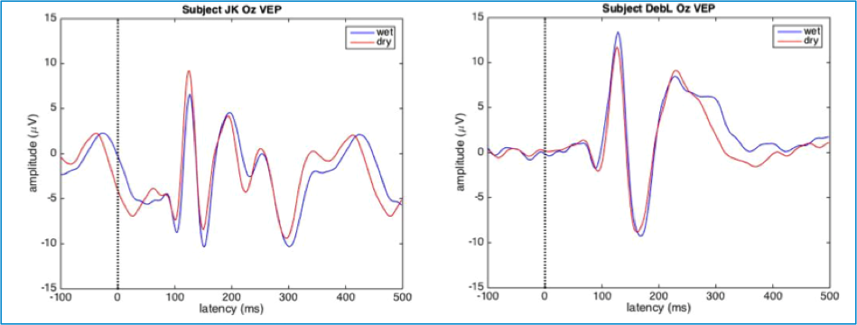
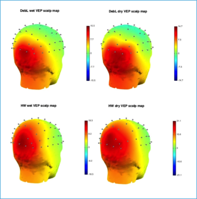
P1 Latency and P1 amplitude
Comparable results were found for conventional gelled and dry electrodes when looking at P1 latency. The observed P1 latency for wet electrodes was 109.7 ms whereas the P1 latency for dry electrodes was found to be 110.2 ms. No statistically significant difference in P1 latency was observed when comparing wet and dry electrodes (p=0.46; two-tailed, paired t-test).
P1 amplitude was also similar for wet and dry cap preparations, with mean values of 11.95 μV and 11.35 μV for wet and dry electrodes, respectively. No statistically significant difference in P1 amplitude could be detected between the wet and dry cases (p=0.29; twotailed, paired t-test).
Rejected electrodes
In this setup, the percentage of trials that were rejected based on visual inspection of the dry EEG signal is roughly 12% higher compared to conventional gelled recording.
Moreover, the percentage of participants in which specific electrodes were rejected is higher when using dry electrodes. This is not uncommon for dry electrodes and is likely due to reasons such as participants’ head shape and hairstyle. The artifact correction module in eego software can be used in dealing with this problem using the SPHARA method. For evoked potentials, many artifact issues are resolved by having a large number of trials.
Impedance levels
There were distinct differences between the impedance thresholds for gelled electrodes and dry electrodes. The impedance levels for conventional gelled electrodes ranged from 1 kΩ to 18 kΩ, whereas impedance for dry electrodes in working mode ranged from 25kΩ to 1MΩ. The signal analysis showed that the dry electrodes can perform well under higher impedances due to the active shielding technology and in combination with the eego
amplifiers.
Summary
In this multicenter study, the waveguard touch with dry electrodes was compared to conventional gelled caps. Electrophysiologically, no statistically significant differences were found between dry and gelled electrodes when comparing important pattern reversal VEP metrics, and qualitatively similar eye blink waveforms, pattern reversal VEPs and resting EEG spectra are observed for both preparations.
Based on these results, we can state that the ANT waveguard touch cap reduces setup time considerably while providing high quality EEG data. It is an excellent alternative for conventional gelled sensors for research purposes.
Related publications
- Fiedler, P., Pedrosa, P., Griebel, S., Fonseca, C., Vaz, F., Supriyanto, E., Zanow, F., Haueisen, J. (2015). Novel multipin electrode cap system for dry electroencephalography. Brain Topography, 28, 647-656
- Fiedler, P., Strohmeier, D., Hunold, A., Griebel, S., Mühle, R., Schreiber, M., Pedrosa, P., Vasconcelos, B., Fonseca, C., Vaz, F. and Haueisen, J., 2016. Modular multipin electrodes for comfortable dry EEG. Presented at the 38th Annual International Conference of the IEEE Engineering in Medicine and Biology Society (EMBC).
- Graichen, U., Eichardt, R., Fiedler, P., Strohmeier, D., Zanow, F., Haueisen, J. (2015). SPHARA - A generalised spatial Fourier analysis for multi-sensor systems with non-uniformly arranged sensors: Application to EEG. PLoS ONE 10(4): e0121741.


ANT Neuro
Welbergweg 74
7556 PE Hengelo
Netherlands
T: +31 (0) 85 049 8175
F: +31 (0) 85 049 3919
E: Send us an email